THE BARD INSTITUTE
Professional Association of Non-Invasive Diagnostic Science
 |
Dr. Robert L. Bard dedicated his life's work to the expansion of diagnostic imaging in the medical community. His undying commitment to public advocacy of safer, non-invasive protocols and a global movement in integrative medicine is what helped to forge The Bard Institute for Non-Invasive Diagnostic Science.
Established in 2020, the principles of this association was originally formed within the many fundamental efforts of the AngioFoundation (2002) and the NY Cancer Resource Alliance (2001). This partnership combined public works encouraging education, research, innovation and the expansion of scientific modalities by innovative thinkers and technical supporters of sustainable health practices. These independent visionaries collectively produced programs and public projects to drive strategic health solutions simply called "the better way".
The core objectives of the Bard Institute include:
- Program 1: Presenting awards and citations to top achievers in advocacy and education
- Program 2: the public promotion of medical grade non-invasive imaging technologies
- Program 3: The establishment and support of educational training courses in utlrasound technology
- Program 4: Building a national coalition of experts in diagnostic imaging
- Program 5: The collaboration of united professionals in pursuit of research and furthering the development of all diagnostic solutions
 |
PART 1:
PROMOTION OF LEADERSHIP & ADVOCACY
A major part of THE BARD INSTITUTE's commitment to social causes is the public support for education, research and technical achievements. Angiofoundation was originally designed to offer scholarships to those in pursuit of higher learning in the field of innovative medicine. In addition, the Bard Institute also supports public programs raising awareness in prevention, early detection and safety by caregivers, public speakers and educators who are committed to public outreach.
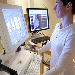
PART 2: ULTRASOUND TRAINING PROGRAM
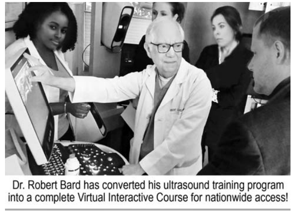 |
For all nursing programs, emergency care training and general field medicine, the future of patient care involves the use of non-invasive Digital Imaging. The faculty of the Bard Institute, comprised of certified radiologists and cancer diagnostic imaging experts is offering a COMPREHENSIVE ULTRASOUND TRAINING PROGRAM and a review of the Point of Care Ultrasound System (POCUS) designed for all areas of health studies. This course work covers the most common applications of scanning protocols including: GENERAL IN-OFFICE SCANNING, RESCUE RESPONSE DIAGNOSTICS, PREOP scanning, IMAGE GUIDED PROCEDURES and POST-OPERATIVE SCANS. Thanks to the digital innovations and web-based collaboration tools of advanced ultrasound solutions, empowering your students’ with the ability to operate the latest point-of-care imaging devices brings them to the next generation of efficiency, performance and professional value.
In addition, our program also offers an additional WORKSHOP IN IMAGING INTERPRETATION- sharing his specialized experience-based review on the methods of correctly assessing and translating the apparent and not-so obvious results of some of the most complex ultrasound scans. A must-have for any health professional.
VIRTUAL COURSE & TELEMED SCANS
This course work is dedicated to ‘empowering’ all health care and medical personnel with a full range of technical and analytical applications of how to properly operate a digital ultrasound scanner. With a combination of our video training materials and real-time (Video Conferencing) instructional guidance, Dr. Bard’s training program will establish the main operating essentials and a full understanding of screen data breakdown, probe uses and selections, anatomic analysis and how and when to implement features such as the Doppler system and Elastography.
PRACTICE EXPANSION COURSEWORK 1: Empowering Your Private Practice with Ultrasound Training
Eliminate “best guess” evaluations and potential surprises. BDI offers a complete training and technology guidance program to support medical imaging for your private practice. With today’s affordable digital innovations, imaging devices have become portable, affordable and more powerful to get more answers about what’s really underneath the patient’s skin. Adding imaging to your health services is a priceless upgrade to identifying your patients’ condition while elevating a new level of confidence, safety and accuracy to your entire treatment process.
TOP BENEFITS OF IN-HOUSE IMAGING & OVERREADING PARTNERSHIP
• Expand your patient diagnostic and reporting services to include medical imaging
• Gain user proficiency in a short amount of time
• Employ a portable scanning system for immediate, real-time patient assessment
• Earn additional peace of mind by preventing possible misreads, oversights or “land mines”
• Enhance your office’s productivity & performance with strong diagnostic support
• Install a turn-key scanning solution with your current office operations and software
• Reduce your patients’ travel time to other locations
• Wifi features allow for outside medical collaboration & remote 2nd opinions
• Improve time management by outsourcing patient analysis to trusted experts
• Implement a streamlined patient reimbursement (direct charging)
• Enable custom reporting of final interpretation
ACCURATE READINGS = MAJOR RISK REDUCTION
Many routine scanning protocols can easily determine the patient's risk level prior to a surgical procedure. Pre-operative imaging is often recommended to verify tissue planes and measure fat depth. DIGITAL PRE-OP is a highly useful prep stage for any medical procedure, preventing potential problems within certain patients who could carry hidden complex issues. Oftentimes, patients may have forgotten prior treatments. A new scan can reveal extensive sub-dermal calcium deposition, unsuspected fluid collections or thick fibrosis distorting the expected anatomy. Anatomic variants may be observed and avoided. Moreover, patient confidence is enhanced by the extra care provided by this advanced technology. But the main advantage for all patients is the experience-based diagnostic assessment combined with the investigative talents of our seasoned imaging readers who are experienced in detecting scanned disorders and issues that many could miss.
PART 3: NETWORKS AND PARTNERSHIPS
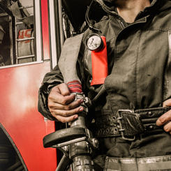
Our outreach connects us with fellow “do-gooder” groups and individuals. Through the power of NETWORKING, SHARING & COLLABORATION, we explore ways where we can align with others’ initiatives or brainstorm to create new ones. We seek out specific target areas who need support (ie. Cancer exposures in rescue service). We aim to do this effectively in various ways:
1) EDUCATION: Many of our medical members are also educators in their own right. Offering PREVENTION, EARLY DETECTION or LIFESTYLE IMPROVEMENT wisdom can come in the form of seminars/webinars, sharing original articles or videos.
2) AWARENESS INITIATIVES: We all have social media platforms, websites, subscribers and followers. Supportive partnership could mean “getting the word out” about you, your current work or any programs you wish to distribute to an audience larger than your own.
3) RESOURCE SHARING: Collectively, we have a massive catalog of resources- which includes professional experts, innovative technologies, facilities etc. For our allies, we welcome the opportunity to help those who seek resources to help anyone in need.
4) CO-PUBLISHING & PROMOTING: We support a growing number of partnerships in publishing, video production and public relations- where ‘getting the word out’ also means re-distributing news and science articles based upon relevance. (One network is www.NYCRANEWS.com)
5) BUILD EXCLUSIVE CLINICAL (DIAGNOSTIC OR TREATMENT) PROGRAMS: Many of our health professionals welcome working with associations (some offer free help, discounted services or other added benefits) and the establishment of an exclusive patient care program specific to groups.
6) TECHNICAL ADVISORY: Many of our professionals (both clinical and non) offer their credentials and actual support on an advisory level and varying capacities.
7) TECHNICAL TRAINING: As educators and clinicians, certain members welcome the opportunity to build support training programs in varying levels. For example, a radiologist may offer remote online training on how to use an ultrasound for ambulance units, triage units or field rescue groups.
8) RESEARCH PROGRAMS: Clinical trials, pilot research projects or product/process testing.
9) THINK TANKS AND POWERMEETS™: For our members who relate with your mission, they can form a collaborative panel to join your group brainstorming session.
Research & Educational PROGRAMS
The AngioFoundation has been recognized worldwide by official medical organizations and peer reviews for its work in FIVE MAIN focal topics and class divisions. We continually maintain and update research works in these categories to support the advancement of these disciplines as part of The AngioFoundation's commitment to the scientific community.
 Advanced use of Doppler Sonographic Imaging technology to identify malignant cancers and monitor their behavior through blood flow parameters- (Breast, Lung, Bladder, Prostate, Melanoma, etc) which correlates with comparative studies with other current technologies such as MRI, CT etc. Advanced use of Doppler Sonographic Imaging technology to identify malignant cancers and monitor their behavior through blood flow parameters- (Breast, Lung, Bladder, Prostate, Melanoma, etc) which correlates with comparative studies with other current technologies such as MRI, CT etc. |
 Research / assessment of musculoskeletal disorders (arthritis, inflammation, trauma) and dermatological issues through the advanced use of 3D/4D ULTRASOUND innovations. Research / assessment of musculoskeletal disorders (arthritis, inflammation, trauma) and dermatological issues through the advanced use of 3D/4D ULTRASOUND innovations. |
 TECH REVIEWER: Beta-testing, industry-wide comparative feature review/evaluation program. Drafting of FDA application / compliance documentation of digital imaging technologies (models, brands and generations) including sub-dermal and musculoskeletal treatment devices TECH REVIEWER: Beta-testing, industry-wide comparative feature review/evaluation program. Drafting of FDA application / compliance documentation of digital imaging technologies (models, brands and generations) including sub-dermal and musculoskeletal treatment devices |
 Internal study of all cancer issues and health disorders of victims associated with 9/11 and other disaster-related environmental toxic exposures. Collaboration with geological labs & environmental statistics. (See First Responders Cancer Resource) Internal study of all cancer issues and health disorders of victims associated with 9/11 and other disaster-related environmental toxic exposures. Collaboration with geological labs & environmental statistics. (See First Responders Cancer Resource) |
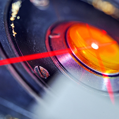 Function review / performance evaluation program of all laser-based medical equipment including devices specializing in sub-dermal musculatory treatment of chronic disorders. Function review / performance evaluation program of all laser-based medical equipment including devices specializing in sub-dermal musculatory treatment of chronic disorders. |
Research results in APPLIED SCIENCE IMAGING ASSISTS SURGICAL PLANNING OF INDICATED BIOPSIES
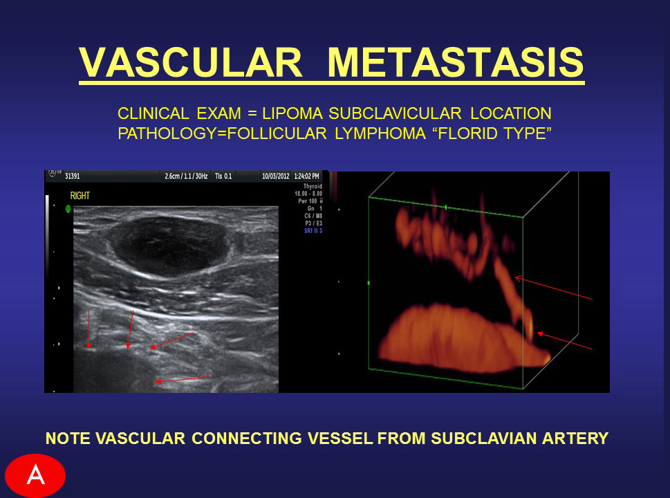
In my extensive career as the medical director of an advanced imaging diagnostics practice, I have provided great assistance to many surgeons with my work using advanced Doppler Scanning of Tumors and Cosmetic Disorders. I have uncovered countless dermal and subcutaneous issues that would have otherwise gone undetected with less effective technologies, leading to potential complications in the surgical procedure and patient recovery. The advancement in this innovation empowers upcoming surgical procedures with remarkable confidence of a safer end result. Where biopsies are becoming a thing of the past, our non-invasive 4D Digital imaging replaces weeks of lab work and radiologic tests and often provides more useful information. DIGITAL BIOPSY CASES: WHAT ARE YOU ABOUT TO BIOPSY? WHAT HAPPENS AFTER THE NEEDLE INSERTS? Here we have 2 subdermal masses which are non tender and firm with no history of trauma. Case A: The oval mass (dark echoes=suspicious) with irregular vessels (red) was referred as a probable cyst or lipoma. The tumor is highly vascular and connected from the aorta by way of the subclavian feeding artery. Liposuction could result in massive hemorrhage and spread of tumor cells into the circulation.
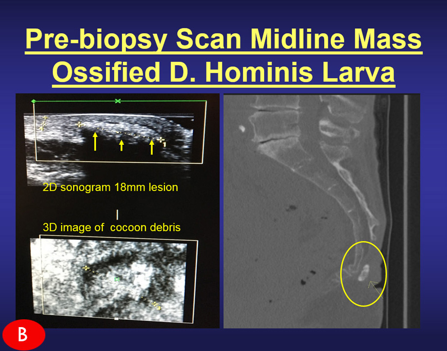
Case B: The ovoid white region (bright echoes=benign) is ossified as confirmed by the CT scan of the coccyx. The sonogram allows you to reassure the patient it is NOT CANCER. It prompts one to avoid a standard needle that could bend, crack or dislodge into the soft tissues requiring further exploration to locate/retrieve the broken metal fragment. |
|||
For more information or to donate to the AngioFoundation, contact us at: 631-920-5757 or email us at: AngioFoundation@gmail.com
|
|||
| ALSO SEE: MommiesOnAMission.org |